
Diagnosis
To diagnose Myasthenia Gravis, the following tests can be done:
- Blood tests
- Neurological examination
- Imaging tests (e.g., x-ray, CT scan)
- Tensilon test
- Neurological tests (e.g., electromyography)
Blood tests 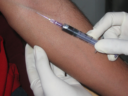
These are performed to determine the serum levels of certain antibodies. For example; AchR-binding antibodies, AchR-modulating antibodies and antistriational antibodies. High levels of these particular antibodies may indicate Myasthenia Gravis.
Image Courtsey of
https://www.flickr.com/photos/fn-goa/169481694/
under a creative commons license
Neurological examination
This involves testing muscles and reflexes. To test arm and leg muscles, the patient may be instructed to maintain a position against resistance for a period of time. Weakness that occurs during this test is called fatigability.
CT scans and Chest x-rays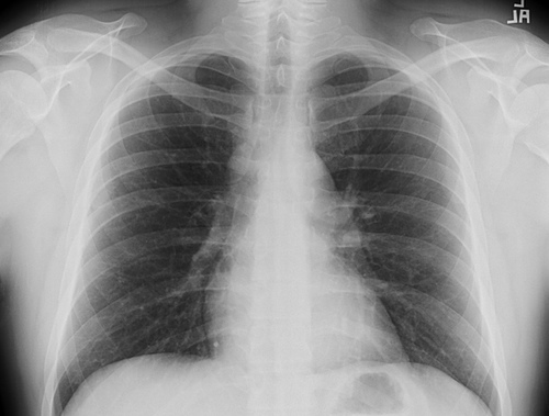
Computed tomography (CT scans) and chest x-rays may be performed to detect an enlarged thymus (thymoma), which is common in MG.
Image Courtesy of
https://www.flickr.com/photos/evanskrig/2141439225/
under a creative commons license
Tensilon test
The is often used to diagnose MG. The enzyme acetylcholinesterase breaks down acetylcholine (ACh) after the muscle is stimulated, preventing prolonged muscle response to a single nerve impulse. Edrophonium chloride (Tensilon) is a drug that temporarily blocks the action of acetylcholinesterase.
In MG, there are few acetylcholine receptor sites (AChR) on the muscle and acetylcholine is broken down before it can fully stimulate the muscle, resulting in muscle weakness. By blocking the action of acetylcholinesterase, Tensilon prolongs muscle stimulation and temporarily improves strength.
In this test, Tensilon is administered intravenously (into a vein) and muscle response is evaluated. In cases of MG, muscle weakness improves within 1 minute. The Tensilon test is most effective when easily observed weakness is present, and is less useful for vague or fluctuating complaints. Side effects of the test include temporary abnormal heart rhythms such as rapid heart rate (atrial fibrillation) and slow heart rate (bradycardia).
Electromyography
Electromyography (EMG) uses electrodes to stimulate muscles and evaluate muscle function. Muscle contractions that become progressively weaker may indicate MG.
Contact
Search site
Did you know...
Dogs and cats can get typical MG, with the usual antibodies that weaken nerve to muscle triggering in human patients. Therefore it is thought that MG must have always been with us.